
Introduction to ESD and EMR
EMR (Endoscopic Mucosal Resection) is a procedure performed under endoscopy to remove flat or elevated lesions, such as early-stage gastrointestinal cancer or flat adenomas. This is achieved by separating the lesion from its inherent layer through injection and suction, creating a pseudopolyp, and then using techniques like snaring or electrocautery. ESD (Endoscopic Submucosal Dissection), on the other hand, is a technique developed based on EMR, which involves the complete removal of the mucosal layer under endoscopy. ESD is primarily used for early-stage digestive tract cancer and precancerous lesions.
Historical Development
The history of endoscopic tumor resection can be traced back to 1974 in Japan. The development of endoscopic tumor resection originated from the use of high-frequency electrical current for colon polypectomy. In 1974, Japan was the first to use endoscopic polypectomy to treat pedunculated or sessile early gastric cancer (EGC). By 1984, a technique called "strip biopsy," which is a form of EMR, was introduced. This technique utilized a dual-channel endoscope and was relatively simple to perform while ensuring a complete histopathological diagnosis of the resected specimen to assess the adequacy of resection. The procedure involved injecting saline solution beneath the lesion's submucosa, lifting the lesion with forceps, and then resecting it using a snare inserted through the second channel. This method gained widespread acceptance in Japan as an endoscopic treatment strategy for small EGC.

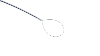
EMR with a transparent cap (EMRC) assisted by a transparent cap was introduced in 1992 for the resection of early esophageal cancer and has gradually been applied to the removal of early gastric cancer (EGC). The characteristic feature of EMRC is the placement of a transparent cap in front of the endoscope lens, with different sizes available depending on the endoscope diameter and lesion size. After submucosal injection at the lesion site, a specially designed crescent-shaped snare is placed in the groove at the top of the transparent cap. By suctioning, the lesion is drawn into the cap and captured and tightened by the snare. As the electrical incision is performed from the submucosal layer, it is considered safe. EMR-L technique utilizes a standard variceal ligator to band the lesion, making it resemble a polyp. The advantages of EMR-C and EMR-L are their relative simplicity and the use of a regular endoscope without the need for additional assistance. However, these techniques are not suitable for complete resection of lesions larger than 2 cm. Since piecemeal resections hinder pathologists from accurately staging and assessing the completeness of resection, and carry a higher risk of recurrence, further methods have been developed for en bloc resection of larger lesions.

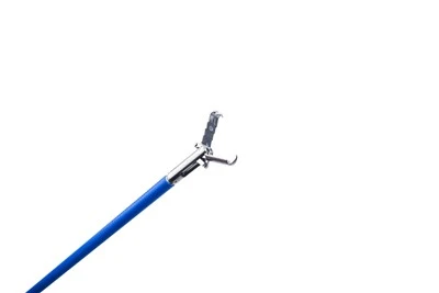
The technique of endoscopic submucosal dissection (ESD), which involves using an improved needle-knife under direct visualization of the submucosal layer, was first introduced by the National Cancer Center Hospital in Japan. The concept of ESD using the IT knife originated from the improvements made in endoscopic resection of early gastric cancer (ERHSE), initially performed by surgeons but now safely and conveniently carried out by endoscopists. ESD offers advantages over standard endoscopic mucosal resection (EMR) as it allows for en bloc resection using a single-channel endoscope, resulting in a complete specimen. ESD makes it possible to remove larger lesions in one piece, achieving more accurate histological staging and preventing recurrence compared to standard EMR methods. Other ESD instruments have gradually emerged, such as the hook knife, flex knife, and small-caliber transparent cap. Despite the higher skill requirements and longer procedure time, ESD has become widely practiced and is considered the preferred method for en bloc resection of larger early gastric cancer lesions.